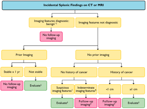
Spleen

†Hemangioma: discontinuous, peripheral, centripetal enhancement (findings that are uncommon in splenic hemangiomas).
‡Benign imaging features: homogeneous, low attenuation (20 HU), no enhancement, smooth margins.
§Evaluate: PET vs. MRI vs. biopsy. Suspicious imaging features: heterogeneous, enhancement, irregular margins, necrosis, splenic parenchymal or vascular invasion, substantial enlargement.
¶Indeterminate imaging features: heterogeneous, intermediate attenuation (20 HU), enhancement, smooth margins.
#Follow-up MRI in 6 and 12 months.
Lymph Nodes

†Abnormalimaging features: enlarged short-axis diameter (1 cm in retroperitoneum), architecture (round, indistinct hilum), enhancement (necrosis/hypervascular), increased number (cluster of 3 lymph nodes in single nodal station or cluster of 2 lymph nodes in 2 regions).
‡Nonneoplastic disease: eg, infection, inflammation, connective tissue.
§Other evaluation (PET/CT, nuclear scintigraphy [MIBG],endoscopic ultrasound).