Table of Contents
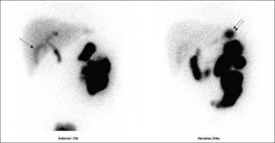
V/Q Scans

NucMedResource: Interpreting VQ scans
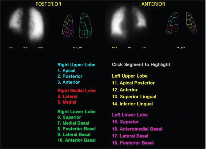
Lung Segments
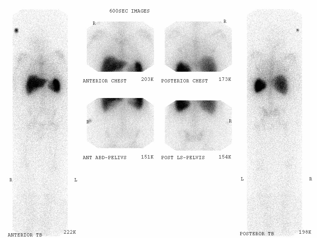
Infection/Inflammation
Gallium normal biodistribution

WBC scan normal biodistribution
HIDA Scan
“The hallmark of acute cholecystitis (acalculous as well as calculous) is persistent gallbladder nonvisualization after 3–4 h of passive imaging or 30 min after morphine administration. A pericholecystic hepatic band of increased activity (rim sign) is a sign of severe late-stage acute cholecystitis and has been associated with severe phlegmonous or gangrenous acute cholecystitis, a surgical emergency.”