Table of Contents
- Intracranial Hemorrhage
- General Anatomy Relevant to Stroke
- Stroke
- Maxillofacial Trauma
- Odontogenic Infection
- Visceral Neck Trauma
- Spinal Trauma
- Programmable Shunt Settings
- Deep Spaces of the Head and Neck
- CT Sinus Anatomy
- Temporal Bone Anatomy
For carotid ultrasound please refer to the ultrasound section.
INTRACRANIAL HEMORRHAGE
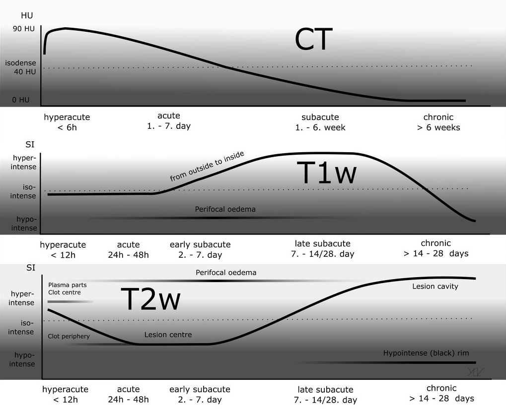
Evolution of Blood Products

GENERAL STROKE ANATOMY
Scrollable Lobar Anatomy
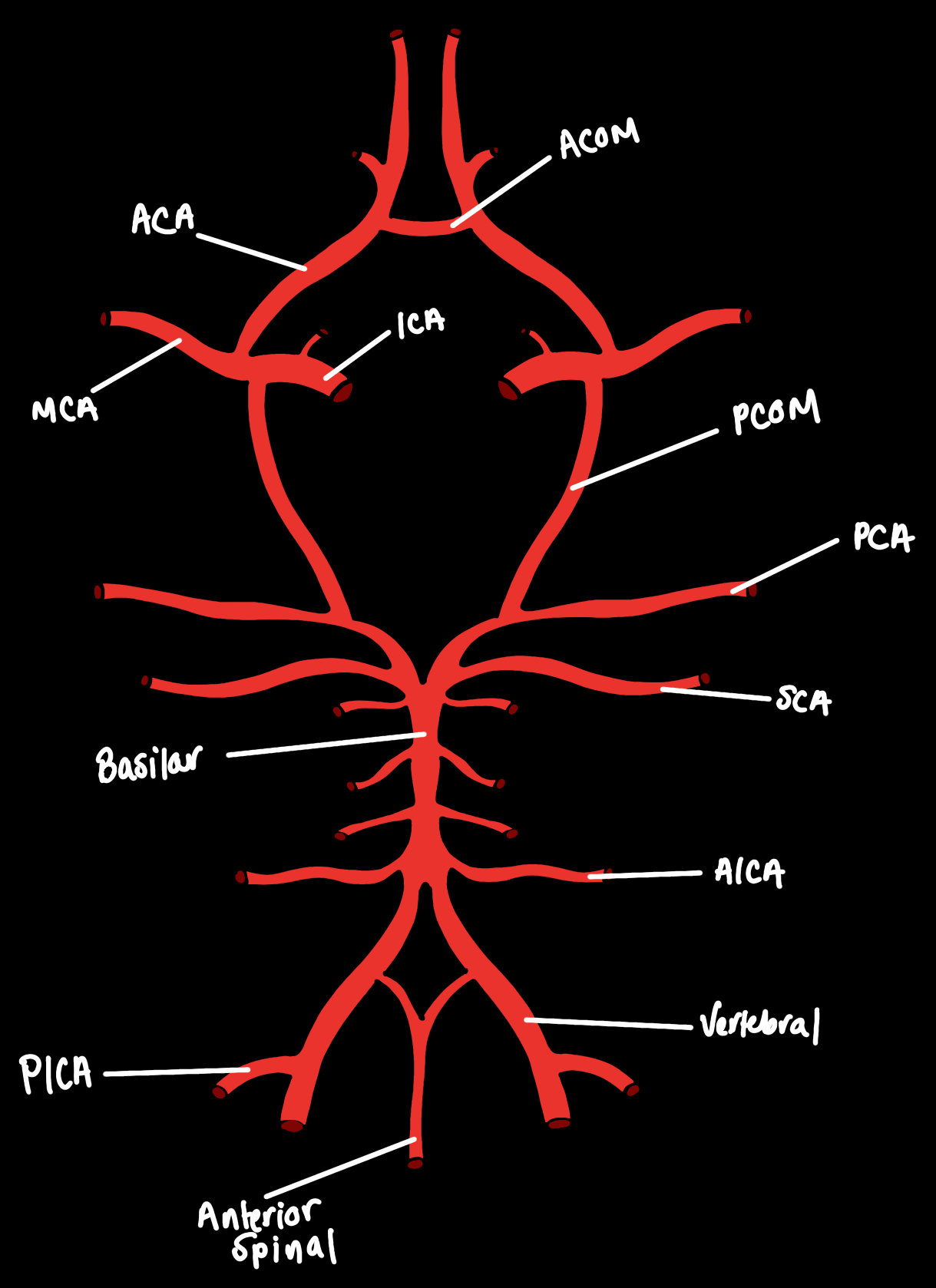
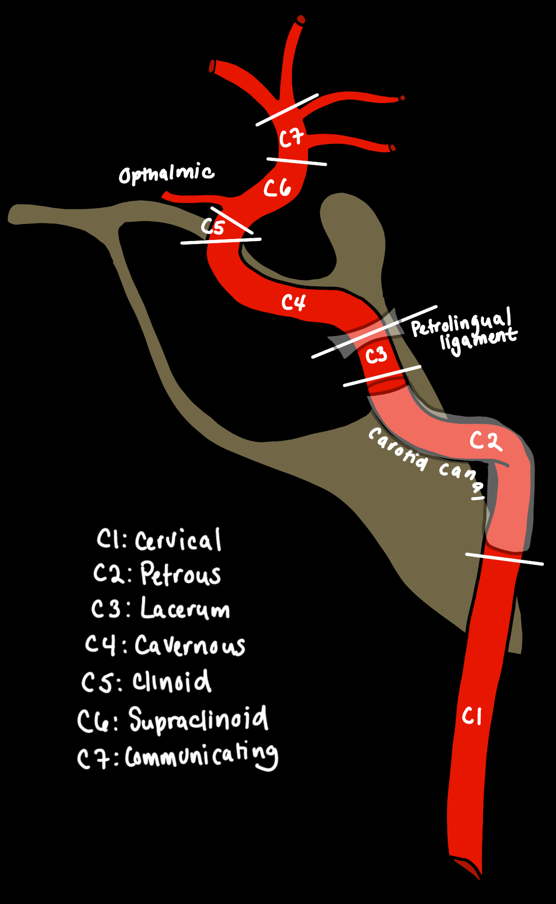
Vascular Anatomy
Vascular Territories
Scrollable Vascular Territories


STROKE
Stroke Quick Reference
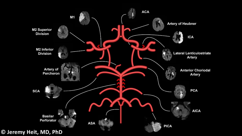
ASPECTS (MCA stroke)
- 1 point for each area involved

CT Perfusion
Thrombectomy (simplified)
- Core infarct < 70mL
- Tissue at risk >15mL
Blunt Cerebrovascular Injury
Denver Criteria
When to screen for BCVI:
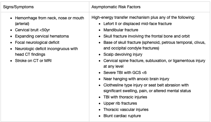
Biffle/Denver Grade of BCVI
| Grade I | luminal irregularity or dissection with <25% luminal narrowing |
| Grade II | dissection or intramural hematoma with ≥25% luminal narrowing, intramural thrombus, or raised intimal flap |
| Grade III | pseudoanuerysm |
| Grade IV | occlusion |
| Grade V | transection with free extravasation |

MAXILLOFACIAL TRAUMA
Overview of Facial Trauma
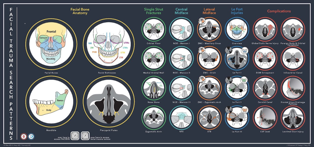


NOE Types

| Type | Description |
| Type I | Large bone fragment |
| Type II | Comminution with fracture lines central to tendon |
| Type III | Central comminution with fracture lines beneath insertion of the tendon |
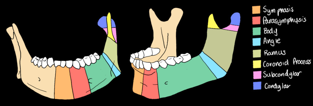
Mandibular Anatomy
ODONTOGENIC INFECTION

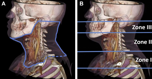
VISCERAL NECK TRAUMA
SPINAL TRAUMA
AO Spinal Injury Classification
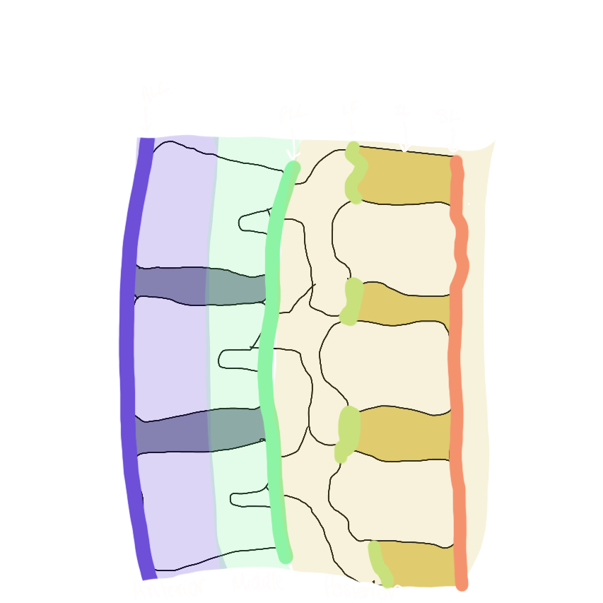
Three column concept of the spine (Denis)
Simplified. If ≥2 contiguous spinal columns are disrupted the fracture will be considered unstable. For the lumbar spine but can be extrapolated to the other segments
Cervical Spine
Basic search pattern for traumatic cervical spine CT

Normal Craniocervical Measurements in Adults

| Basion-dens interval | < 9.5mm |
| Powers ratio | < 0.9mm |
| Atlanto-dental interval | <3.0mm |
| Atlanto-occipital interval | <1.4mm |
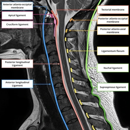
Ligamentous Anatomy of the Cervical Spine

Atlanto-Occipital Dissociation
- Basion-Dens interval >9.5mm
- Alar ligament injury, tectorial membrane disruption
- Associated injuries
- SAH at foramen magnum
- Avulsion of the basion, tip of the dens, or occipital condyles
Occipital Condyle Fractures
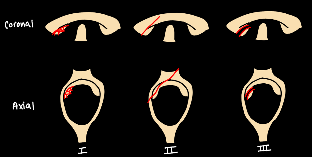
| Type | Characteristics |
| Type 1 | Comminuted, impacted |
| Type 2 | Extension of skull fracture |
| Type 3 | Disruption of Alar ligament (unstable) |
C1-C2 Injury
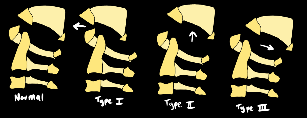
Atlantoaxial Rotary Fixation
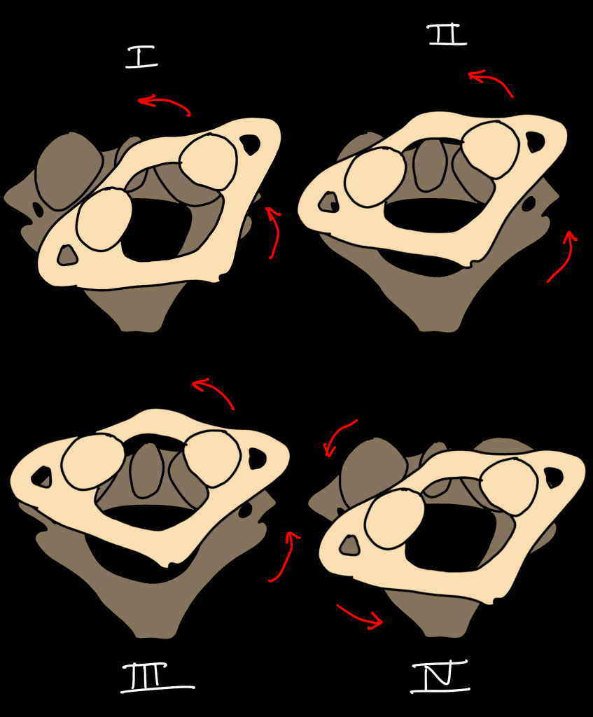
| Type | Characteristics |
| Type I | Intact transverse and alar ligaments |
| Type II | Transverse ligament disruption; 2-3mm anterior translation with rotation |
| Type III | Alar and transverse ligaments disrupted; >5mm anterior translation with rotation |
| Type IV | Alar and transverse ligaments disrupted; anterior displacement of C1 over C2 with deficient odontoid |
Atlas Fractures

- Look for the bilateral displacement of the lateral masses on coronal views.
- Burst and lateral mass fractures can be associated with tears of the transverse ligament, which are unstable.
- Rule of Spence – separation of fracture fragments >5.4mm on CT = transverse ligament disruption and instability
- Ruptures of the ligament can compromise the atlantodental relationship, causing dorsal displacement of the dens, and possibly resulting in compression of the thecal sac.
- Jefferson Fracture = C1 burst fracture
Odontoid Fractures

| Type | Characteristics |
| Type I | Apical avulsion fracture (stable in isolation) |
| Type II | Transverse fracture – most common and unstable. If >4mm displacement or comminuted risk nonunion |
| Type III | Extends into the C2 body; unstable |
Hanged Man Fracture
- Hyperextension injury with fracture of both pars interarticularis or pedicles of C2
- Atypical fracture – propegation into the vertebral body – get CTA

| Type | Characteristics | Management |
| Type I | Bilateral pars fractures without translation or angulation | Stable; rigid collar |
| Type II | >3mm anterior displacement or significant angulation C2/3 disc disruption and PLL disruption Most common | Unstable, reduction or surgery depending on the degree of displacement |
| Type IIa | Significant angulation without anterior displacement Fracture line is more horizontal | |
| Type III | Anterior translation and angulation with facet subluxation or dislocation | Unstable, surgery |
Subaxial Spine
- Injury is most common around C4/C5
Hyperflexion Injury
- Progressive ligamentous injury from posterior to anterior
- Interspinous process widening
- Uncovering of facet joints
- Widening of the posterior disc space – unstable
- Focal kyphosis
- Anterior subluxation
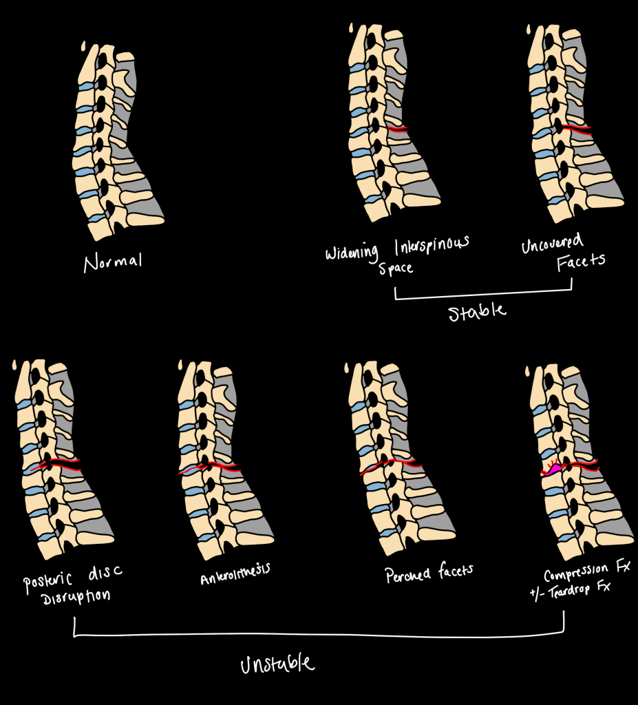
- Hyperflexion sparain – isolated PLC disruption – stable
- Anterior subluxation – disruption of the PLV and posterior annulus – unstable (2 column injury)
- Interfacetal dislocation – unstable
- Perched or jumped facets
- Bilateral or unilateral
- All should undergo MRI
- Flexion teardrop – most severe hyperflexion injury
- Anterior cord syndrome – qudriplegia with pain and temperature insensitvity, preserved vibratory and position sense
Hyperextension Injuries
- Can be easy to miss
- Hyperextension dislocation – disruption of all three columns and unstable
- Anterior disc space widening
- Facet widening
- Extension teardrop fracture

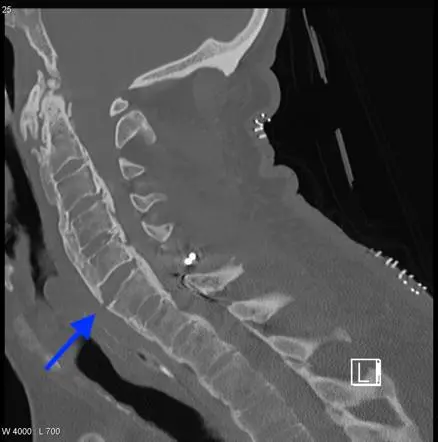
Fused Spine Injury
- Chalk stick type fractures – hairline fracture through a fused spine, can be easy to miss
- Seen in hyperflexion and hyperextention injury
- If seen – image the entire spine as noncontiguous fractures are common
Thoracolumbar Spine
Injury pattern is based on AO Classification which uses the tension band concept
- Anterior Tension Band – limits extension
- Posterior Tension Band – limited flexion

AO Spine Classification for Thoracolumbar Injuries
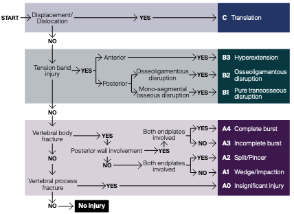
Compression Fracture (A1)
- Failure of the anterior column
- Isolated anterior compression fracture is considered stable
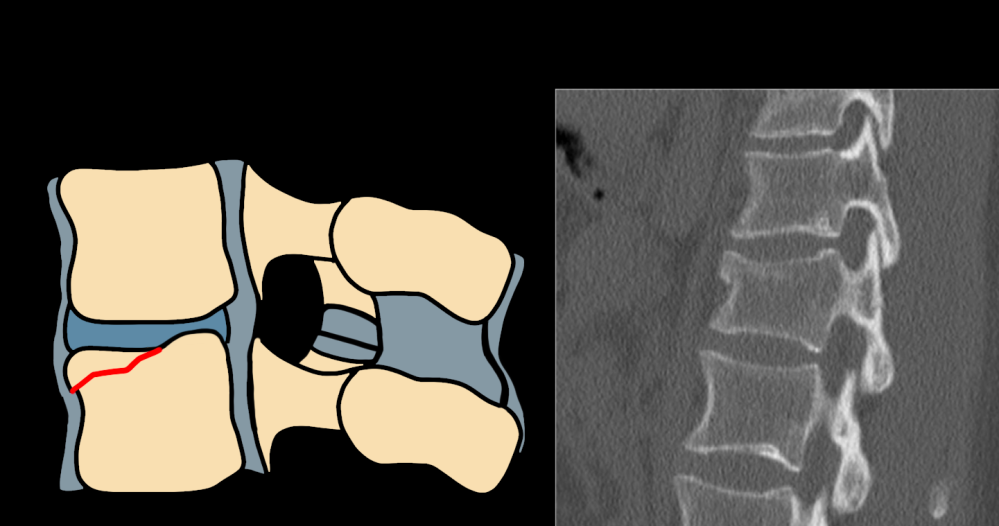
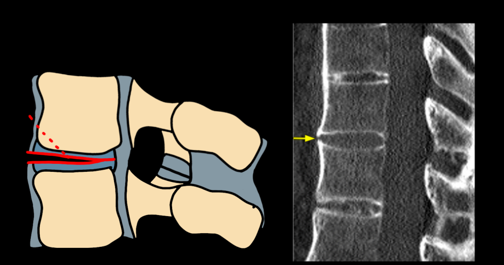
Pincer Fracture (A2)
- Complete coronal split fracture with central collapse and herniation of disc material into the fracture
- Unstable

Burst Fracture (A3 or A4)
- Disruption of the posterior vertebral body cortex differentiates from compression fracture
- If unclear, look for widening of the interpedicular distance on coronal
- Evalute for retropulsed fragment that can cause cord compression
- Incomplete burst – one endplate involved (A3)
- Complete burst – both endplates invovled (A4)
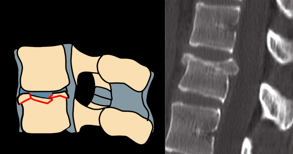
Flexion Distraction Injuries (B1 or B2)
- Transosseous Tension Band Disruption (B1) – Chance Fracture – extends through all through osseous spinal columns
- High association with intraabdominal injuries
- Can also have a pure ligamentous injury – look for distraction and widening of the interspinous space
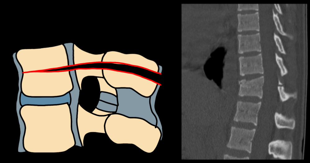
- Ligamentous disruption of the posterior tension band with an class A fracture (B2)
Hyperextension Injury (B3)
- Anterior disc space widening
- Can be seen within rigid spine (see Carrot stick fracture above)
- Can be ligamentous or osseoligamentous
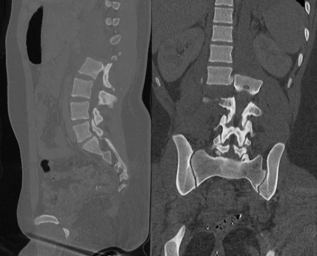
Fracture Dislocation (Type C)
- Disruption of all three spinal columns with distraction, dislocation, or translation
- If you see two vertebral bodies on the same axial image = fracture dislocation
Source: Radiopaedia
