Table of Contents
- Acute abdomen in the neonate
- Atlas of normal measurements in pediatric radiology
- Bones normal for age
- Developmental dysplasia of the hip
- Intraventricular hemorrhage grading
- Lines and tubes in the neonate
- Malrotation
- Neonatal chest radiograph
- Non-accidental trauma
- Normal cranio-cervical measurements pediatrics
- Normal ultrasound measurements in pediatrics
- Pediatric hydronephrosis grading
- Salter Harris classification
- Slipped capital femoral epiphysis
Acute Abdomen in the Neonate
Atlas of Normal Measurements in Pediatric Radiology
Bones Normal For Age
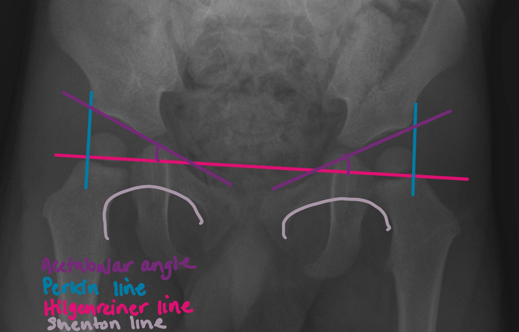
DDH
Alpha angle – should be <60 degrees

Intraventricular Hemorrhage Grading

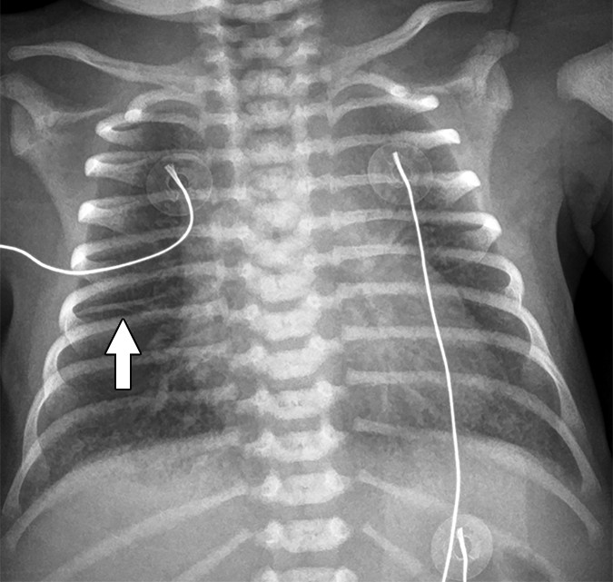
Lines and Tubes
Umbilical artery catheter
- High position: T6-T9 (preferred)
- Low position: L3-L5
- Anywhere else is abnormal

Umbilical venous catheter
- In IVC at the level of the diaphragm

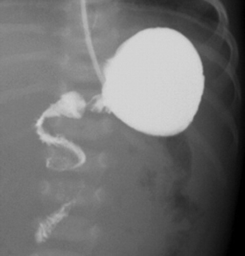
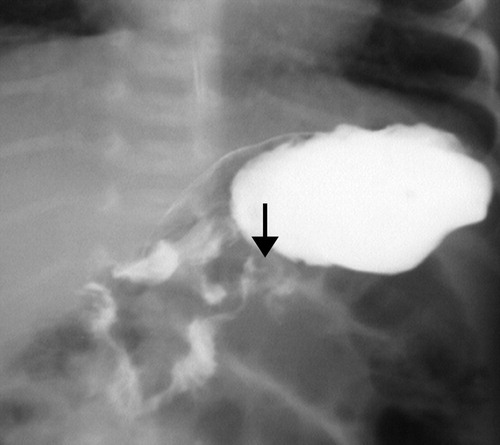
Malrotation

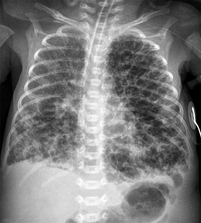
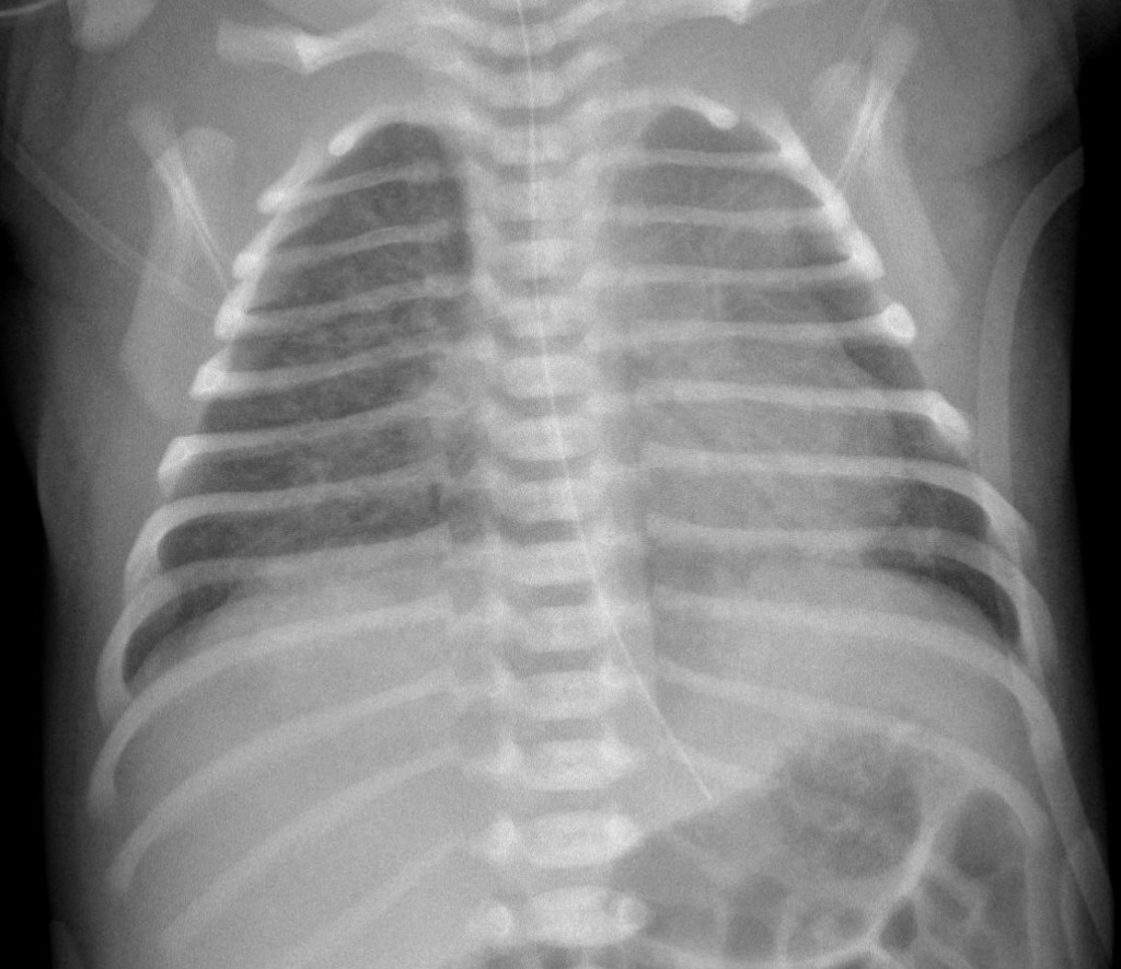
Neonatal Chest Radiograph


Non-Accidental Trauma
- Metaphyseal fractures
- Posterior rib fractures
- Depressed skull fractures
- Multiple fractures of different ages
- Scapular fractures
- Outer third of clavicle fractures
- Sternum fractures
Normal Cranio-Cervical Measurements

| Basion-dens interval | < 7.5mm |
| C2 prevertebral soft tissue | < 5.4mm |
| Atlanto-dental interval | <2.8mm |
| Atlanto-occipital interval | <3.2mm |
| Lateral mass interval | <3.9mm |
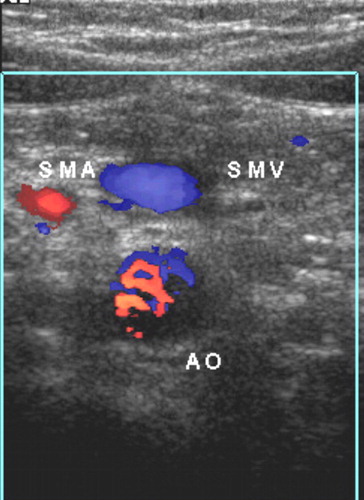
Normal Pediatric Ultrasound Measurements
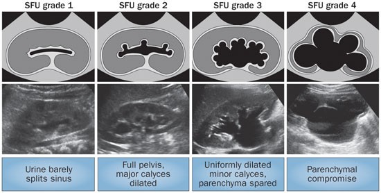
Pediatric Hydronephrosis SFU Grading
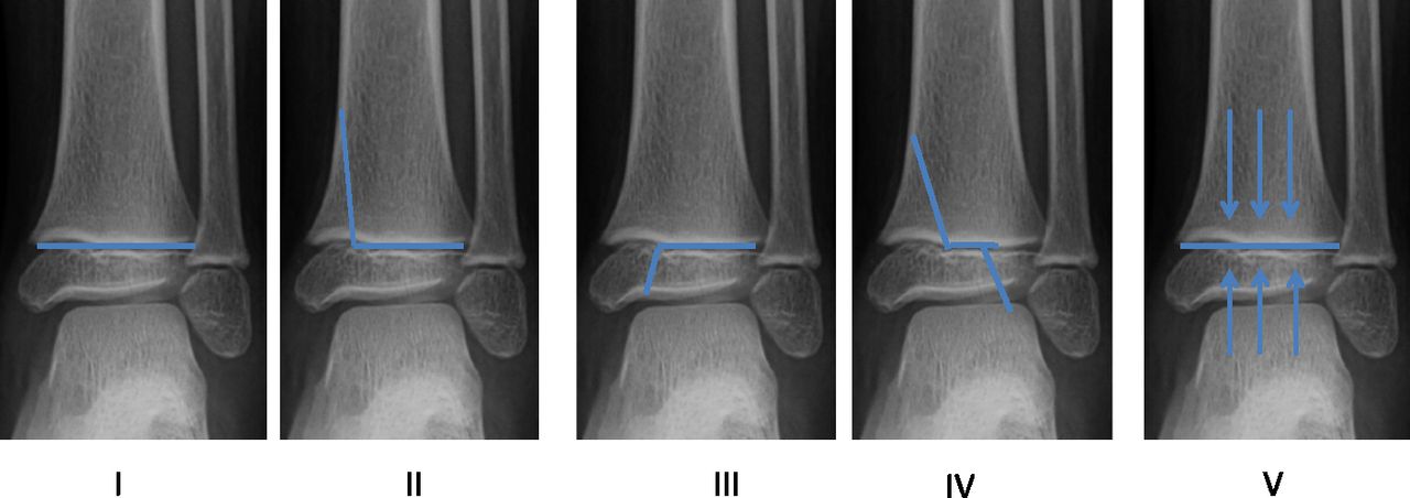
Salter Harris Classification
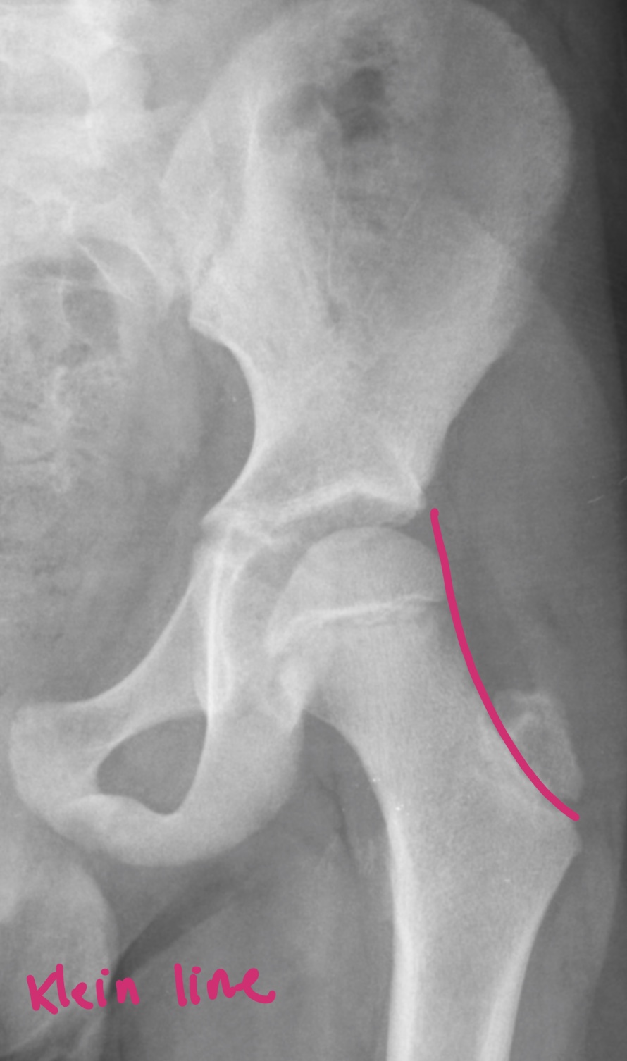
SCFE
Klein’s line – lateral epiphysis should intersect the line. If not, early SCFE
| Mild | lateral edge of epiphysis is within the lateral third of the metaphysis |
| Moderate | lateral edge of epiphysis is within the middle third of the metaphysis |
| Severe | lateral edge of epiphysis is within the medial third of the metaphysis |
Hilgenreiner and Perkin lines
- Hilgenreiner line drawn horizontal through the inferior aspect of both triradiate cartiladge
- Perkin line perpendicular to the Hilgenreiner line, at the lateral most aspect of the acetabulum
- Triradiate cartilage should lie within the upper medial quadrant of where the lines intersect
Acetabular Angle
- Should be <30 degrees at birth and reduce over time