Renal Artery Angioplasty and Stent — Renovascular HTN and FMD
Renal artery revascularization treats hemodynamically significant renovascular disease — atherosclerotic renal artery stenosis (ARAS) in limited clinical scenarios after the CORAL trial, and fibromuscular dysplasia (FMD) in which angioplasty without stenting remains first-line. Successful outcomes depend on rigorous patient selection, a no-touch technique to minimize cholesterol embolization, and careful use of embolic protection.
Key points
CORAL reframed ARAS management: in patients with atherosclerotic RAS and controlled hypertension/stable renal function, optimal medical therapy is equivalent to stenting plus medical therapy — routine stenting for asymptomatic or well-controlled ARAS is not supported.
Current ARAS stenting niches: flash pulmonary edema (Pickering syndrome), acute kidney injury after RAAS blockade, rapidly declining renal function, resistant hypertension on ≥3 drugs, or hemodynamically significant stenosis with objective ischemia evidence.
FMD is a primary interventional indication: plain balloon angioplasty (PTA) alone is first-line for medial fibroplasia — >80% clinical improvement; stent reserved for bailout (dissection, recoil) only.
Significant stenosis = >70% angiographic diameter reduction OR 50–70% with a resting translesional peak systolic gradient >20 mmHg or Pd/Pa <0.9.
Flash pulmonary edema with bilateral RAS or RAS to solitary kidney
Pickering syndrome; strong clinical indication — CORAL excluded these patients
Acute kidney injury after ACEI/ARB initiation
Bilateral RAS functionally unmasked; revascularization preserves renal function
Rapidly declining renal function with hemodynamically significant RAS
Especially if function deteriorates over weeks to months; ischemic nephropathy
Resistant hypertension
On ≥3 antihypertensives (including diuretic) at maximum tolerated doses; hemodynamically significant stenosis
Fibromuscular dysplasia with hypertension
Primary intervention with PTA alone; best in young women with new-onset HTN
Transplant renal artery stenosis
Increasing creatinine, rising HTN; PTA ± stent
Renal artery aneurysm or dissection
Covered stent for select cases
Type
Contraindication
Absolute
Non-viable kidney (length <7 cm, resistive index >0.80, cortical thinning) · Uncorrectable coagulopathy · Active infection · Severe contrast allergy not premedicable
Relative
Well-controlled hypertension on stable regimen with stable renal function (CORAL cohort — medical therapy alone) · Severe CKD (stage 4–5) where contrast risk outweighs benefit · Diffuse atherosclerosis of aorta and renal with high embolization risk
Pathology and Diagnostic Criteria
Pathology
Characteristics
Atherosclerotic RAS (~90%)
Ostial or proximal (within 1 cm of origin); older patients, diffuse atherosclerosis; often ostial plaque continuous with aortic atheroma
Fibromuscular dysplasia (~10%)
Mid to distal main renal artery and branches; "string of beads" on angiography in medial fibroplasia (most common subtype); young to middle-aged women
Kidney length: viable >8 cm; poor recovery if <7 cm or resistive index >0.80
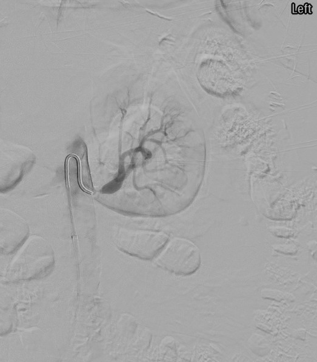
Selective angiogram demonstrating severe left renal artery stenosis — high-grade ostial narrowing consistent with atherosclerotic renal artery stenosis.
CORAL in context: CORAL (2014, NEJM) randomized 947 patients with atherosclerotic RAS plus HTN or CKD to optimal medical therapy with or without stenting. No significant difference in cardiovascular/renal events at ~3.6 years. Key caveats: CORAL excluded flash pulmonary edema, very rapid GFR decline, and single-kidney RAS — revascularization remains appropriate in these high-risk subsets. Patient selection has shifted dramatically toward objective hemodynamic and clinical indicators.
Procedure Overview
The following is a high-level summary. Full catheter and sheath selection, stent sizing matrices, pressure-gradient measurement technique, and embolic protection device selection are available in RadCall Pro.
Pre-Procedure
Dual antiplatelet therapy: aspirin 81 mg daily plus clopidogrel 75 mg daily (load 300–600 mg if not pretreated) starting ≥3 days pre-procedure when possible.
Hydration: IV isotonic saline before and after; hold ACEI/ARB periprocedurally; avoid metformin around contrast.
Baseline labs: creatinine, potassium, coagulation; renal duplex for comparison.
CO₂ angiography may be used to minimize iodinated contrast in patients with severe CKD.
Access and Diagnostic Angiography
Access: right common femoral artery with 6 Fr sheath (7 Fr if guide-in-sheath needed); brachial or radial alternative for caudally angulated renal arteries.
Aortogram: AP and/or 15–20° LAO; confirm renal artery origins, stenosis location and severity, kidney size.
Position guide catheter in aorta adjacent to but not engaging the ostium.
Advance a 0.035" J-wire through the guide catheter into the aorta as a leading wire to hold the guide away from the ostial plaque.
Advance a 0.014–0.018" coronary-style guidewire alongside the J-wire through the guide into the renal artery, crossing the lesion into a distal segmental branch.
Advance the guide catheter over the coronary wire into the renal ostium — the J-wire prevents direct engagement with plaque, minimizing atheroembolism.
Angioplasty and Stenting
Pressure-gradient measurement (if indeterminate 50–70% stenosis) with pressure wire; confirm hemodynamic significance before intervention.
Primary PTA for FMD: high-pressure balloon sized 1:1 with reference vessel; multiple inflations; stent only for bailout (dissection, elastic recoil).
Primary stenting for atherosclerotic ostial RAS: balloon-expandable stent sized to reference vessel diameter (typically 5–7 mm); deploy with 1–2 mm protrusion into aorta to ensure ostial coverage; post-dilate to optimize apposition.
Embolic protection device (filter-type) in select high-risk lesions (severe stenosis, ulcerated plaque, long lesion, solitary kidney).
Completion angiogram: residual stenosis <30%, no dissection, no distal embolization, preserved branch perfusion. Repeat pressure gradient if used.
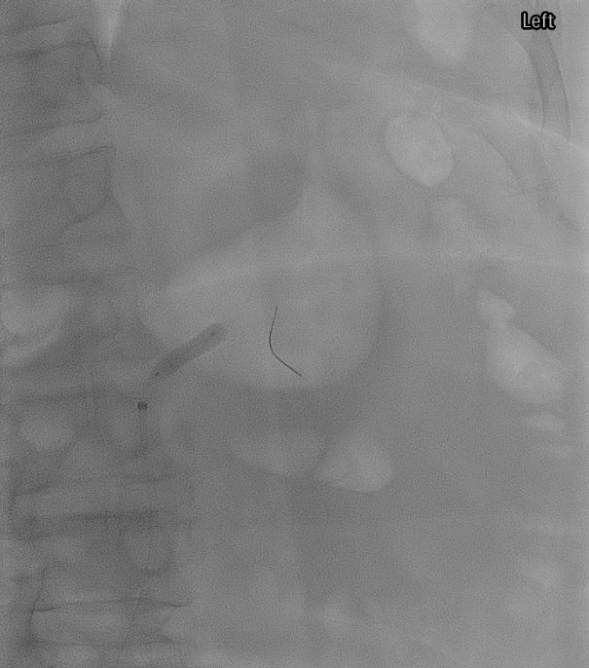
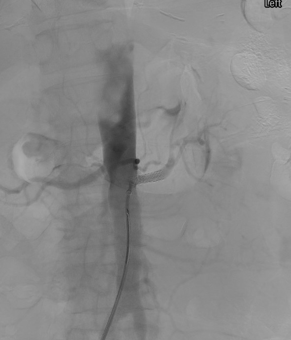
Left renal artery balloon angioplasty (PTA) — balloon inflated across the stenosis prior to stent deployment.Left renal artery stent placement — balloon-expandable stent deployed across the ostial stenosis with 1–2 mm protrusion into the aorta to ensure complete ostial coverage.
Complications
Complication
Rate
Management
Cholesterol / atheroembolization
1–10%
Livedo reticularis, eosinophilia, worsening renal function, blue-toe; supportive; prevented by no-touch and embolic protection
Contrast-induced nephropathy
5–15% in CKD
Hydration; minimize contrast; CO₂ adjunct
Renal artery dissection
1–3%
Bailout stent for flow-limiting dissection; conservative for small spiral dissection
Renal artery perforation / rupture
<1%
Balloon tamponade; covered stent; emergent surgery
Access-site hematoma / pseudoaneurysm
1–3%
Compression, thrombin injection, or US-guided therapy
In-stent restenosis
10–20% at 1 year
Duplex surveillance; re-intervention (PTA ± drug-coated balloon) if hemodynamically significant
Stent thrombosis
<1%
Dual antiplatelet therapy prevents
Segmental renal infarction
1–3%
From embolization to a polar or accessory branch; usually asymptomatic
Post-Procedure Care
Monitoring: overnight observation; vital signs, access site, creatinine day 1.
Antiplatelets: dual antiplatelet therapy for 1–3 months (aspirin + clopidogrel), then aspirin indefinitely.
Blood pressure: expect lability; reduce antihypertensives gradually guided by response.
Follow-up: renal duplex at 1, 6, 12 months and annually; creatinine and BP tracking.
Restenosis management: re-angiography if BP or renal function worsens; repeat intervention if hemodynamically significant.
Evidence Summary
CORAL (Cooper CJ et al, 2014, NEJM): 947 patients with atherosclerotic RAS randomized to stent + OMT vs OMT alone — no difference in composite cardiovascular and renal events at ~3.6 years. Established optimal medical therapy as default for stable ARAS.
ASTRAL (Wheatley K et al, 2009, NEJM): 806 patients — no significant benefit of revascularization for atherosclerotic RAS on renal function; frequent procedural complications.
STAR (Bax L et al, 2009, Ann Intern Med): similar negative findings for stenting in ARAS with impaired renal function.
Kadian-Dodov D et al (2016, J Am Coll Cardiol, US Registry for FMD): PTA alone achieves excellent clinical response in FMD; stent rarely indicated.
Gornik HL et al (2019, JSVS / FMD guidance): diagnostic and treatment framework for FMD.
Ritchie J et al (2014, Am J Kidney Dis): high-risk subsets (flash pulmonary edema, rapid function decline) benefit from revascularization despite CORAL-era conservatism.
References
Cooper CJ, Murphy TP, Cutlip DE, et al. Stenting and medical therapy for atherosclerotic renal-artery stenosis. N Engl J Med. 2014;370(1):13–22.
ASTRAL Investigators. Revascularization versus medical therapy for renal-artery stenosis. N Engl J Med. 2009;361(20):1953–1962.
Bax L, Woittiez AJ, Kouwenberg HJ, et al. Stent placement in patients with atherosclerotic renal artery stenosis and impaired renal function: a randomized trial. Ann Intern Med. 2009;150(12):840–848.
Olin JW, Gornik HL, Bacharach JM, et al. Fibromuscular dysplasia: state of the science and critical unanswered questions: a scientific statement from the American Heart Association. Circulation. 2014;129(9):1048–1078.
Kadian-Dodov D, Gornik HL, Gu X, et al. Dissection and aneurysm in patients with fibromuscular dysplasia: findings from the U.S. Registry for FMD. J Am Coll Cardiol. 2016;68(2):176–185.
Ritchie J, Green D, Chrysochou C, Chalmers N, Foley RN, Kalra PA. High-risk clinical presentations in atherosclerotic renovascular disease: prognosis and response to renal artery revascularization. Am J Kidney Dis. 2014;63(2):186–197.
Weinberg I, Keyes MJ, Giri J, et al. Blood pressure response to renal artery stenting in 901 patients from five prospective multicenter FDA-approved trials. Catheter Cardiovasc Interv. 2014;83(4):603–609.
Full technique in RadCall Pro
Catheter and sheath selection, stent sizing matrices, translesional pressure gradient technique, embolic protection device selection, and periprocedural medication protocols available in RadCall Pro.