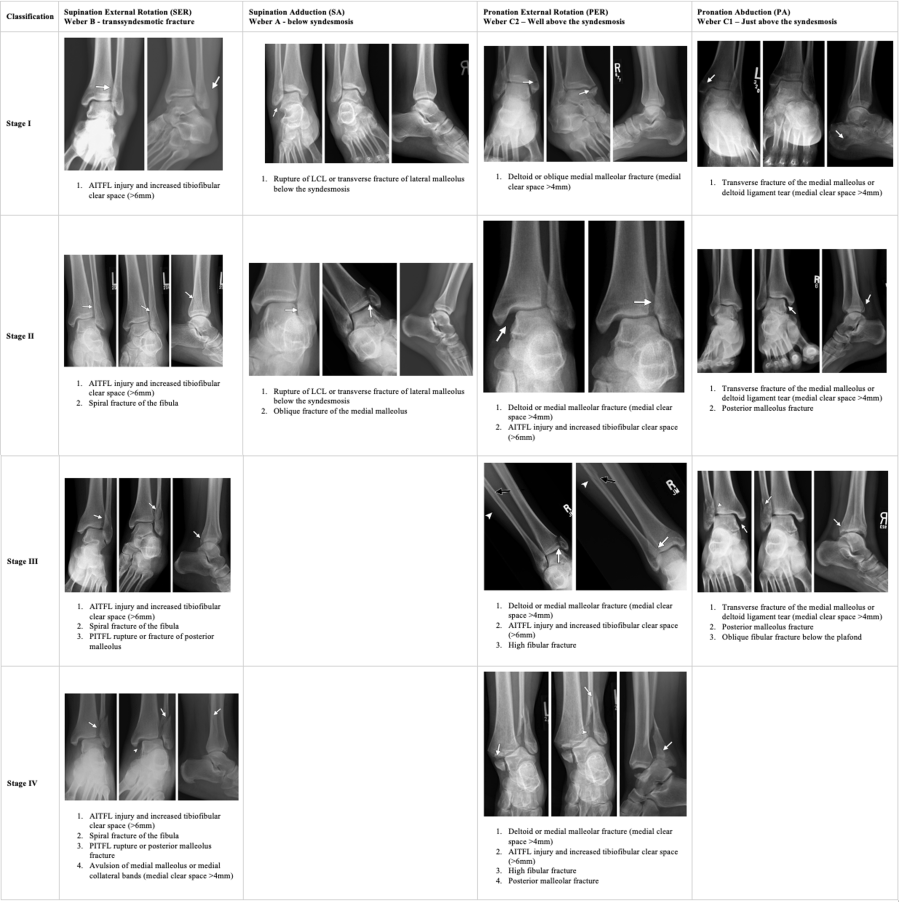
Lauge-Hansen Classification
The Lauge-Hansen system classifies ankle fractures by foot position at time of injury plus direction of the deforming force. The first word describes foot position; the second describes the direction of force. Fractures progress through sequential stages — later stages imply all prior stages are present.
Supination-Adduction (SA)
| Stage | Injury |
|---|---|
| I | Transverse fibula fracture at or below the joint |
| II | Vertical medial malleolus fracture |
Supination-External Rotation (SER)
Most common ankle fracture pattern.
| Stage | Injury |
|---|---|
| I | Anterior tibiofibular ligament tear |
| II | Spiral or oblique fibula fracture at joint level |
| III | Posterior malleolus (posterior tibiofibular ligament) |
| IV | Medial malleolus fracture or deltoid ligament tear |
Pronation-Abduction (PA)
| Stage | Injury |
|---|---|
| I | Medial malleolus fracture or deltoid ligament tear |
| II | Anterior and posterior tibiofibular ligament disruption |
| III | Comminuted fibula fracture at or above joint |
Pronation-External Rotation (PER)
| Stage | Injury |
|---|---|
| I | Medial malleolus fracture or deltoid ligament tear |
| II | Anterior tibiofibular ligament disruption |
| III | Interosseous membrane disruption |
| IV | Spiral fibula fracture well above the joint (Maisonneuve) |
Weber Classification
The Weber system classifies ankle fractures by the level of the fibula fracture relative to the ankle mortise.
| Weber Type | Fibula Level | Syndesmosis | Stability |
|---|---|---|---|
| A | Below mortise | Usually intact | Usually stable |
| B | At mortise | Variable | Depends on medial side |
| C | Above mortise | Disrupted | Unstable |
Syndesmotic Assessment
Lateral clear space greater than 4 mm indicates syndesmotic disruption. Evaluate both the lateral and medial clear spaces for ligamentous integrity.
Maisonneuve fracture: Proximal fibula fracture associated with medial ankle injury and interosseous membrane disruption — always image the full tibia and fibula when medial ankle injury is present without a distal fibula fracture.
Reporting Checklist — Ankle
- Fracture level and pattern (spiral, oblique, transverse, comminuted)
- Weber type and Lauge-Hansen pattern; look for missed associated injuries
- Lateral clear space (>4 mm = syndesmotic disruption)
- Medial clear space (ligamentous integrity)
- Dislocation of the tibiotalar joint
- Syndesmotic disruption; bony fragments within the syndesmosis
- Posterior malleolus involvement (size, articular step-off)
- Maisonneuve fracture: if suspected, image full tibia/fibula
Tibial Plafond (Pilon) Fractures — Ruedi-Allgower
Pilon fractures are high-energy axial-load fractures of the distal tibial articular surface (plafond). Mechanism is axial compression — fall from height or motor vehicle collision. Treatment is typically staged: initial spanning external fixator followed by delayed ORIF.
| Type | Description |
|---|---|
| I | Cleavage fracture without articular displacement — undisplaced |
| II | Displaced fracture with minimal comminution |
| III | Significant comminution with articular impaction |
Reporting Checklist — Pilon
- Number and location of major articular surface fragments
- Degree and location of articular depression (>2–3 mm is significant)
- Syndesmotic disruption; bony fragments within the syndesmosis
- Associated tendon injury or entrapment
- Retinaculum injury
Reference
Okanobo H et al. Simplified diagnostic algorithm for Lauge-Hansen classification. RadioGraphics. 2012;32(2):E71–84.