Column Anatomy
The acetabulum is described using a two-column concept:
- Anterior column: pelvic brim, anterior wall, superior pubic ramus, anterior iliac wing
- Posterior column: greater sciatic notch, posterior wall, ischial tuberosity
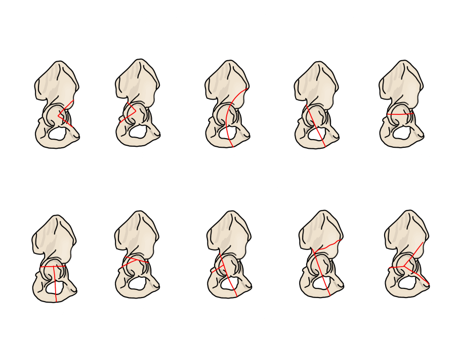
Judet-Letournel Classification
Elementary Fractures
Elementary fractures involve a single anatomic element.
| Pattern | Notes |
|---|---|
| Anterior wall | Involves the anterior rim without the column |
| Anterior column | Extends from iliac crest to superior pubic ramus |
| Posterior wall | Most common elementary fracture; surgical if >40% involvement or with instability |
| Posterior column | Involves the greater sciatic notch through the ischial tuberosity |
| Transverse | Single horizontal fracture line dividing the acetabulum into superior and inferior halves |
Associated Fractures
Associated fractures represent combinations of elementary patterns.
| Pattern | Notes |
|---|---|
| T-type | Transverse fracture with a vertical limb separating the ischiopubic segment |
| Anterior column + posterior hemitransverse | Anterior column fracture combined with a partial transverse component |
| Both column | Most complex pattern — no articular surface remains connected to the axial skeleton; "spur sign" on obturator-oblique view is pathognomonic |
| Transverse + posterior wall | Most common associated pattern |
Key Concepts
Posterior wall involvement: Surgical intervention is indicated when >40% of the posterior wall is involved or when the hip is unstable. Percentage involvement is estimated on CT axial images.
Articular step-off: The typical surgical threshold is >2–3 mm. Measure on CT.
Both-column fracture: The "spur sign" on obturator-oblique radiograph — a bony prominence of the intact posterior ilium — is pathognomonic and indicates that no articular surface connects to the axial skeleton.
CT requirement: CT is required for all acetabular fractures. It characterizes the fracture pattern, quantifies articular step-off, and identifies intraarticular fragments and marginal impaction not visible on plain film.
Reporting Checklist
- Elementary fracture type: anterior wall / anterior column / posterior wall / posterior column / transverse
- Associated fracture pattern: T-type / anterior column + posterior hemitransverse / both column / transverse + posterior wall
- Femoral head: congruent / subluxed / dislocated (direction)
- Articular step-off: measure in mm (surgical threshold typically >2–3 mm)
- Intraarticular fragments: present / absent; size and location
- Posterior wall deficiency: estimate % involvement (surgical if >40%)
- Marginal impaction of articular surface: present / absent
- Ipsilateral femoral neck fracture: present / absent
Reference
Scheinfeld MH et al. Acetabular fractures. RadioGraphics. 2015;35(2):555–577.