Indications / Contraindications
Indications
- Axial spinal pain (cervical, thoracic, lumbar) attributed to facet arthropathy — characteristically worse with extension and rotation; without significant radicular component
- Diagnostic medial branch block (MBB) to confirm facet origin before radiofrequency ablation (RFA)
- Therapeutic intra-articular injection — short-to-medium term pain relief
- Failed conservative management ≥ 6 weeks
- Clinical criteria favoring facet origin: age >50, no radiation below the knee, pain not worsened with walking, relieved with sitting
Contraindications
- Absolute: Active spinal infection · Uncorrectable coagulopathy · Allergy to contrast or corticosteroids
- Relative: Anticoagulation (hold per SIR Cat 1 guidelines before proceeding) · Uncontrolled diabetes (steroid-related glucose spike) · Prior RFA at same level without adequate washout
- Note: SIR Category 1 — routine coagulation correction not required; targeted hold for therapeutic anticoagulation
Pre-Procedure Checklist
Relevant Anatomy
Facet Joint
- Zygapophyseal joint — articulation between the superior articular process of the vertebra below and the inferior articular process of the vertebra above
- True synovial joint with joint capsule; capacity 1–2 mL — overfilling causes capsular rupture and epidural spread
- Lumbar facets oriented sagittally (resist rotation); cervical facets oriented obliquely
- On oblique fluoroscopy: "Scottie dog" — ear = superior articular process (SAP), eye = pedicle, nose = transverse process, front leg = inferior articular process
Medial Branch Nerve Targets
- Each facet joint is innervated by the medial branch of the dorsal ramus from TWO levels — the level above and the level of the joint itself
- Lumbar L1–L4 medial branch: crosses at junction of transverse process and superior articular process — target is the "waist" or "neck" of the Scottie dog
- L5 dorsal ramus: courses in groove between sacral ala and S1 SAP — slightly different target than L1–L4
- Cervical medial branch: targeted at midpoint of articular pillar (C3–C7)
- Rule: To block a facet at level X, block the medial branches at level X and X-1
Technique
Default RadCall approach · share your own below
Supplies
Steps
Position + fluoroscopy setup
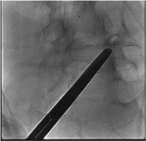
Skin entry + needle advance
Confirm position AP + lateral
Contrast arthrogram

Inject medication
Steps
Identify target levels
AP fluoroscopy — needle to target
Lateral view depth confirmation
Contrast check
Inject local anesthetic ± steroid
Troubleshooting
Intravascular contrast uptake on test injection
Likely cause: Needle tip in epidural venous plexus or periforaminal vessel — more common at cervical and thoracic levels.
Next step: Withdraw needle 1–2 mm and re-aspirate. Redirect slightly. Repeat contrast injection under live fluoroscopy before any medication. If vascular uptake persists, abort and reposition needle. Never inject particulate steroid with active vascular uptake.
Cannot access joint — too narrow or degenerated
Likely cause: Advanced facet arthropathy with complete joint space obliteration; osteophytes blocking entry.
Next step: Increase oblique angle. Try inferior recess first (often most patent). If still inaccessible, target medial branch block instead — equally effective for diagnostic purposes. Consider CT guidance for C7-T1 or severely degenerated joints.
Contrast spreads widely / epidural pattern instead of intra-articular
Likely cause: Needle tip through posterior joint capsule into epidural or paraspinal space; or existing capsular defect from prior injections.
Next step: Withdraw needle to re-enter joint proper. If a capsular defect allows medication to track epidurally, reduce volume to 1 mL total. Document the finding — epidural spread may still be therapeutic but the procedure is technically a pericapsular injection.
Difficult C7-T1 access
Likely cause: Shoulder overlap blocks oblique fluoroscopic view at the cervicothoracic junction.
Next step: Use CT guidance for C7-T1 and T1-2. Alternatively, use a "swimmer's" lateral fluoroscopic view. Pull shoulders caudally with traction during AP/oblique views. Consider prone lateral approach under CT.
Complications
Immediate / Periprocedural
- Post-injection flare (10–20%) — transient pain increase 1–3 days post; resolves spontaneously; ice/NSAIDs helpful; warn patient before discharge
- Intravascular steroid injection — particulate steroid into radicular artery → spinal cord infarct (catastrophic but rare); use live fluoroscopy + contrast mandatory before every injection
- Epidural spread (inadvertent) — from capsular defect or excessive volume; monitor for lower extremity weakness or sensory changes before discharge
- Vasovagal reaction — common; supine positioning, fluids, atropine if needed
- Nerve root injury — rare; usually transient paresthesias from needle contact; reposition immediately
Delayed
- Infection / Septic arthritis (<0.1%) — fever, worsening pain, elevated WBC; spine MRI with contrast; broad-spectrum antibiotics; IR or surgical drainage if abscess forms
- Corticosteroid side effects — hyperglycemia (warn diabetics, 24-48h monitoring), adrenal suppression (limit to 3 injections per region per year), Cushing features with repeated use
- Skin/subcutaneous atrophy — from superficial steroid deposition; use deep, confirmed intra-articular injection
- Hematoma — rare (Cat 1 procedure); usually self-limited
Post-Procedure Care
Same Day
- Observe 20–30 min — check for inadvertent epidural spread (leg weakness) and vasovagal recovery
- Pain diary: instruct patient to document degree and duration of relief starting immediately post-procedure using the online pain diary tool
- Patient must have a driver if local anesthetic was used near neural structures
- Ice application to injection site for discomfort
- Resume normal activity as tolerated — light activity day of, no strenuous exercise for 24h
- Diabetic patients: check blood glucose at home for 24–48h
Follow-up + Decision Points
- Pain diary review at 2 weeks. Document: degree of relief (%), duration of relief (hours/days)
- Positive diagnostic MBB: ≥50–80% pain relief for ≥6 hours with short-acting agent → proceed to RFA workup
- Dual diagnostic blocks: Two positive MBBs (lidocaine then bupivacaine, separate visits) before RFA improves specificity and reduces RFA failure rates
- Therapeutic injection assessment at 4–6 weeks. If >50% relief — document and consider repeat if relapse. If inadequate — reassess diagnosis; consider alternative source (SI joint, disc, myofascial)
- Steroid limit: Maximum 3 injections per anatomic region per year
Critical Pearls
References & Medial Branch Level Guide
Medial Branch Nerve Supply by Facet Level
| Facet Joint | Medial Branch Block Targets | Special Notes |
|---|---|---|
| C3-4 | C3 medial branch (deep) + C4 medial branch — articular pillar midpoint | Third occipital nerve for C2-3; 3rd occipital headache pattern |
| C4-5 | C4 + C5 medial branches | Articular pillar midpoint; lateral approach preferred |
| C5-6 | C5 + C6 medial branches | Most common cervical level treated |
| C6-7 | C6 + C7 medial branches | C7-T1 access often requires CT guidance |
| L1-2 to L3-4 | Level above + same level medial branches (e.g., L3-4: block L2 + L3 MB) | Junction of TP and SAP ("waist of Scottie dog") |
| L4-5 | L3 medial branch + L4 medial branch | Most commonly treated lumbar level |
| L5-S1 | L4 medial branch + L5 dorsal ramus | L5 DR target: sacral ala / S1 SAP groove — different from other levels |
Citations
- Prologo JD, Ray CE Jr., eds. Advanced Pain Management in Interventional Radiology: A Case-Based Approach. Thieme; 2024. DOI: 10.1055/b000000387
- Filippiadis DK, Kelekis A. A review of current trends in spinal interventional procedures for the management of chronic spinal pain. Quant Imaging Med Surg. 2017;7(6):651–659.
- Manchikanti L, et al. Comprehensive evidence-based guidelines for facet joint interventions in the management of chronic spinal pain. Pain Physician. 2020;23(3):S1–S127.
- Bogduk N. International Spine Intervention Society. Practice Guidelines for Spinal Diagnostic and Treatment Procedures. 2nd ed. ISIS; 2013.
- SIR Standards of Practice Committee. Consensus Guidelines for Periprocedural Management of Coagulation Status. J Vasc Interv Radiol. 2012;23(6):727–736.
References & Resources
Key Guidelines
- ISIS Practice Guidelines for Facet Injection
- ASIPP Evidence-Based Guidelines for Diagnostic and Therapeutic Facet Joint Interventions
- ACR Appropriateness Criteria for Low Back Pain
Primary References
- Bogduk N. International Spinal Injection Society guidelines for the performance of spinal injection procedures. Part 1: Zygapophysial joint blocks. Clin J Pain. 1997;13(4):285-302.
- Manchikanti L et al. A systematic review of the effectiveness of lumbar facet joint nerve blocks. Pain Physician. 2007;10(3):425-460.
- Schwarzer AC et al. The prevalence and clinical features of internal disc disruption in patients with chronic low back pain. Spine. 1995;20(17):1878-1883.